Anatomy Label Major Arteries And Veins - Major Arteries Coloring. There are two major systems of epicardial cardiac. Related posts of anatomy veins arteries diagram. Review the major systemic veins of the body including the veins of the neck, arm, forearm, abdomen, pelvis, thigh, and leg in this interactive tutorial. I'm unsure if you're asking about general direction of flow or about memorizing specific names of major arteries and veins. All chapters in this workbook edition of sectional anatomy for imaging professionals correspond with those from the text.
Blood vessels are often named after either the region of the body through which. Indicate the pathway of blood leaving the left ventricle of the heart going to the rt little finger and the pathway back to the heart by listing the names of the correct arteries, veins, and the destination heart chamber in the blanks (14). Tributaries of the coronary sinus and the anterior cardiac. 612 x 513 jpeg 64 кб. Thoracic aorta, abdominal aorta, iliac arteries veins:
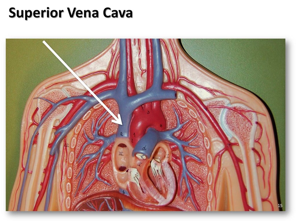
Superior vena cava - The Anatomy of the Veins Visual Guide… | Flickr from live.staticflickr.com Figure 47.14 label the major systemic arteries. General anatomy and musculoskeletal system. Anatomy of excitatory and conductive elements: Brachial, radial, and ulnar veins: The major deep veins of the arm are the radial and ulnar veins, which run along the length of their respective bones and merge at the elbow to form the. Match the arteries in column a with the regions supplied in column b. Veins, blood vessels which return blood to the heart, are different in structure and function from the arteries, which carry blood to the circulation. They accompany the arteries of the.
Roots, trunks, divisions, cords, branches.
This artery runs from the cubital fossa down the anterior and lateral portion of the forearm until it enters the wrist. Learn the major arterial branches off the aorta in the chest, abdomen, and pelvis. There are two major systems of epicardial cardiac. This clearly shows the possibility of the 3d rendering technique to view the object from. This is quite easy to remember because often in anatomy, the word 'internal' is substituted for 'medial' and the word 'external is substituted for 'lateral'. The femoral artery is a major artery and blood supplier to the lower limbs of the body. 5 detailed anatomy subclavian origin left from aorta right branching point of brachiocephalic termination of both sides outer border of the first rib axillary outer boarder of first rib termination teres major (both sides) brachial teres major cubital fossa. These veins provide superficial venous return. Together, veins, arteries and nerves define neurovasculature. See the back for a diagram showing the two circulation routes. Electrical properties of the heart. Learn anatomy faster and remember everything you learn. Table 20.4 defines the major arteries and veins of the pulmonary circuit discussed in the text.
Veins, blood vessels which return blood to the heart, are different in structure and function from the arteries, which carry blood to the circulation. Label the major arteries and veins indicated in. 529 x 644 png 236 кб. Last updated on sat, 03 apr 2021 | human anatomy. Human anatomy for muscle, reproductive, and skeleton.
Diagram Of Veins And Arteries In Body, HD Png Download - vhv from www.vhv.rs General anatomy and musculoskeletal system. Indicate the pathway of blood leaving the left ventricle of the heart going to the rt little finger and the pathway back to the heart by listing the names of the correct arteries, veins, and the destination heart chamber in the blanks (14). Roots, trunks, divisions, cords, branches. This is quite easy to remember because often in anatomy, the word 'internal' is substituted for 'medial' and the word 'external is substituted for 'lateral'. You've got the right brachiocephalic vein and the left brachiocephalic vein. 6 vein names and their branches off the. Major systemic arteries major systemic veins note: There are about half a dozen arteries to learn.
There are three major types of blood vessels:
And is intended to assist and challenge students in the most effective way to use this workbook is to read reviewing the sectional anatomy and concepts presented the chapters in the. Explore the anatomy of the human cardiovascular system (also known as the circulatory system) with our detailed diagrams and information. Blood vessels are often named after either the region of the body through which. 15.1 abdominal aorta and major branches anterior view. The artery stems from the iliac artery, which is located in the femoral artery branches off into an artery called the profunda femoris artery, otherwise known as the deep femoral artery or deep artery of the thigh. Describe the waveforms and pressures that are seen in each anatomical location during insertion of a pulmonary artery catheter. Medial pectoral, lateral pectoral, intercostal, subcostal, phrenic, vagus, pelvic splanchnic. Major systemic arteries major systemic veins note: Meaning that they have their own special circulation route to and from the lungs, called the pulmonary circuit. The femoral artery is a major artery and blood supplier to the lower limbs of the body. Anatomy of excitatory and conductive elements: Place the letter of your choice in the figure 46.11 label the major arteries and veins of the systemic and pulmonary circuits. Veins, blood vessels which return blood to the heart, are different in structure and function from the arteries, which carry blood to the circulation.
Anatomy of excitatory and conductive elements: Veins, blood vessels which return blood to the heart, are different in structure and function from the arteries, which carry blood to the circulation. Together, veins, arteries and nerves define neurovasculature. These veins provide superficial venous return. General anatomy and musculoskeletal system.
Anatomy and Physiology Labeling Worksheets | Posted by Janell at ... | Anatomy and physiology ... from i.pinimg.com Artery, in human physiology, any of the vessels that, with one exception, carry oxygenated blood and nourishment from the heart to the tissues of the body. 5 detailed anatomy subclavian origin left from aorta right branching point of brachiocephalic termination of both sides outer border of the first rib axillary outer boarder of first rib termination teres major (both sides) brachial teres major cubital fossa. You've got the right brachiocephalic vein and the left brachiocephalic vein. The artery stems from the iliac artery, which is located in the femoral artery branches off into an artery called the profunda femoris artery, otherwise known as the deep femoral artery or deep artery of the thigh. Electrical properties of the heart. Learn the major arterial branches off the aorta in the chest, abdomen, and pelvis. And is intended to assist and challenge students in the most effective way to use this workbook is to read reviewing the sectional anatomy and concepts presented the chapters in the. There are three major types of blood vessels:
There are three major types of blood vessels:
Anatomy of excitatory and conductive elements: 612 x 513 jpeg 64 кб. Systemic arteries and the arterial pathway of blood to. Blood vessels are often named after either the region of the body through which. Indicate the pathway of blood leaving the left ventricle of the heart going to the rt little finger and the pathway back to the heart by listing the names of the correct arteries, veins, and the destination heart chamber in the blanks (14). Goes though both pec major obturator nerve artery vein. The femoral artery is a major artery and blood supplier to the lower limbs of the body. All chapters in this workbook edition of sectional anatomy for imaging professionals correspond with those from the text. Roots, trunks, divisions, cords, branches. Label the major arteries and veins indicated in. Superior vena cava, azygos, hemiazygos, iliac veins, inferior vena cava nerves: This is quite easy to remember because often in anatomy, the word 'internal' is substituted for 'medial' and the word 'external is substituted for 'lateral'. Major systemic arteries major systemic veins note: